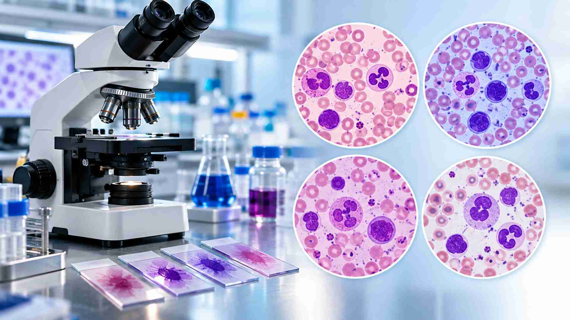
In hematology and diagnostic microscopy, selecting the right stain is essential for obtaining clear, interpretable cellular details. Romanowsky stains are a group of laboratory stains widely used to differentiate blood cells, identify cellular structures, and support the examination of blood and tissue samples. Among the most commonly used options are Wright’s, Giemsa, Leishman’s, and Jenner’s stains. Although they share similar staining principles, differences in composition and application make each one suitable for specific laboratory requirements.
Understanding these differences helps laboratory professionals, researchers, and chemical buyers select an appropriate stain for routine blood smear examination, parasite detection, cytology, or specialized research.
What Are Romanowsky Stains?
Romanowsky staining is based on the interaction between acidic and basic dyes. Traditional formulations generally contain eosin, an acidic dye that stains basic cellular components in shades of pink or red, and methylene blue or its oxidation products, which stain acidic structures such as nuclei and certain cytoplasmic components in blue to purple shades.
The characteristic purple coloration of nuclear chromatin and other cellular structures results from the interaction of these dye components. This contrast makes it easier to distinguish different blood cell types and observe their morphology under a microscope.
Wright’s Stain: A Practical Choice for Blood Smears
Wright’s stain is a widely used Romanowsky-type stain in hematology. It typically contains eosin Y and methylene blue derivatives dissolved in methanol. The methanol acts as both a solvent and a fixative, helping preserve cellular morphology during staining.
Common applications
- Peripheral blood smear examination
- Differential white blood cell counting
- Evaluation of red blood cell morphology
- Examination of bone marrow smears
- General hematological investigations
Advantages and limitations
Wright’s stain provides useful contrast between erythrocytes, leukocytes, and platelets, making it suitable for routine hematology. It is relatively convenient because fixation and staining can occur in the same process.
However, staining results can vary with reagent quality, buffer conditions, and staining time. Some applications requiring particularly strong nuclear or parasite detail may benefit from other formulations or modified staining procedures.
Giemsa Stain: Detailed Cellular and Parasite Visualization
Giemsa stain is another important Romanowsky stain, commonly associated with detailed staining of blood cells and microorganisms. It contains azure dyes, methylene blue, and eosin, typically prepared in a methanol-based formulation. Giemsa is often used as a diluted working solution in buffered water.
Common applications
- Detection and examination of blood parasites
- Malaria parasite microscopy
- Examination of blood and bone marrow smears
- Cytogenetic and cytological preparations
- Identification of cellular and microbial structures
Advantages and limitations
Giemsa stain is valued for its ability to produce strong nuclear detail and useful contrast in cellular preparations. It is particularly important in parasitology, where staining can help reveal parasite morphology in blood films.
Its performance depends heavily on dilution, buffer pH, staining duration, and the type of specimen. Laboratories must use validated procedures because inappropriate conditions may produce excessive background staining or obscure important structures.
Leishman’s Stain: Commonly Used in Routine Hematology
Leishman’s stain is a Romanowsky-type stain frequently used for peripheral blood film examination and differential leukocyte counting. It generally contains eosin and methylene blue or related azure compounds in methanol.
Common applications
- Peripheral blood smear staining
- Differential leukocyte counting
- Examination of blood cell morphology
- Routine hematology laboratories
- General diagnostic microscopy
Advantages and limitations
Leishman’s stain is popular in routine hematology because it combines fixation and staining in a methanolic solution. It can provide clear differentiation of leukocyte nuclei, cytoplasm, and granules when used correctly.
However, the quality of the result depends on factors such as smear thickness, stain concentration, buffer pH, and washing technique. It may not be the first choice for every specialized application, particularly when a laboratory requires a specific staining protocol for parasite identification or cytogenetic analysis.
Jenner’s Stain: A Simpler Romanowsky Formulation
Jenner’s stain is an earlier Romanowsky-type formulation used primarily for blood and cellular staining. It generally contains eosin and methylene blue in methanol. Compared with some other Romanowsky formulations, Jenner’s stain is often described as having a simpler dye composition.
Common applications
- Blood smear examination
- General hematological staining
- Demonstration of blood cell morphology
- Laboratory and teaching microscopy
Advantages and limitations
Jenner’s stain can be useful for routine cellular staining and provides contrast between different blood cell components. Its methanolic formulation also supports fixation during the staining process.
However, it may provide less extensive differentiation than formulations containing a broader range of azure dye components. The choice between Jenner’s and other Romanowsky stains should therefore depend on the intended application and the level of cellular detail required.
Wright’s vs Giemsa vs Leishman’s vs Jenner’s Stain
| Feature | Wright’s Stain | Giemsa Stain | Leishman’s Stain | Jenner’s Stain |
| General composition | Eosin and methylene blue derivatives in methanol | Eosin, methylene blue, and azure dyes | Eosin and methylene blue or related azure compounds in methanol | Eosin and methylene blue in methanol |
| Primary applications | Routine hematology and blood smears | Blood parasites, cytology, and detailed cellular examination | Peripheral blood films and differential counts | General blood cell staining |
| Key strength | Practical blood cell differentiation | Strong nuclear and parasite detail | Convenient routine hematology staining | Simpler formulation for cellular staining |
| Important considerations | Buffer and staining time affect results | Dilution and pH are particularly important | Smear quality and staining conditions matter | May offer less differentiation for specialized applications |
These distinctions are general, as commercial formulations and laboratory methods may differ.
How to Choose the Right Romanowsky Stain
The most suitable stain depends on the specimen, diagnostic objective, and laboratory workflow.
- For routine peripheral blood smears – Wright’s or Leishman’s stain may be appropriate where the primary goal is blood cell differentiation and differential leukocyte counting.
- For blood parasite examination – Giemsa stain is widely used, particularly when parasite morphology and cellular detail are important.
- For general teaching or cellular morphology – Jenner’s stain may be suitable for basic blood cell staining applications.
- For research and specialized microscopy – Consider the required level of nuclear, cytoplasmic, or microorganism detail, along with the laboratory’s validated staining protocol.
Conclusion
Wright’s, Giemsa, Leishman’s, and Jenner’s stains are important Romanowsky-type laboratory stains with overlapping but distinct applications. Wright’s and Leishman’s stains are commonly associated with routine hematology, Giemsa is particularly valuable for parasite and detailed cellular examination, and Jenner’s offers a simpler formulation for general blood cell staining.
The right choice depends on the specimen, staining objective, and laboratory protocol. By understanding the differences between these Romanowsky stains, laboratory professionals and chemical buyers can make more informed decisions and achieve reliable, reproducible staining results.