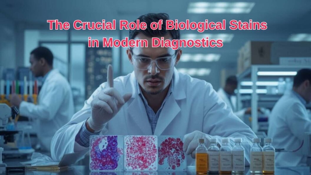
Modern medicine relies heavily on precision, and nowhere is this more evident than in diagnostics. Whether it’s identifying bacterial infections, studying cell structures, or diagnosing cancer, biological stains play a pivotal role in making the invisible visible. These stains transform transparent specimens into vibrant, detailed visuals, allowing pathologists and researchers to detect abnormalities with remarkable accuracy.
In the age of advanced molecular diagnostics and digital imaging, biological stains remain indispensable. They form the foundation of microscopic analysis, bridging the gap between science and visual interpretation. Companies like GSP Chem, a leading name in the specialty chemicals and reagent industry, continue to refine and supply these essential compounds to laboratories worldwide, ensuring reliability, purity, and consistency in diagnostics.
What Are Biological Stains?
Biological stains are colored chemical compounds used to highlight specific components within biological samples like tissues, cells, or microorganisms. When applied, they bind to particular cell structures such as nuclei, cytoplasm, or cell walls, enabling scientists to visualize different elements under a microscope.
Depending on the purpose, stains can be classified as:
- Basic Stains (e.g., Methylene Blue, Crystal Violet): Bind to acidic components like DNA and RNA.
- Acidic Stains (e.g., Eosin, Picric Acid): Attach to basic structures such as cytoplasm and collagen.
- Neutral Stains (e.g., Romanowsky-type stains): Provide balanced coloration, ideal for blood smears and cytology.
In diagnostics, each stain serves a unique role, ensuring accuracy in identifying cellular morphology, bacterial types, and tissue characteristics.
Historical Perspective: From Simple Dyes to Diagnostic Tools
The use of biological stains dates back to the late 19th century, when scientists like Robert Koch and Hans Christian Gram pioneered staining techniques to identify pathogens.
- Gram’s Stain (1884) – Differentiated bacteria into Gram-positive and Gram-negative groups, a practice still central to microbiology today.
- Hematoxylin and Eosin (H&E) – Became the cornerstone of histology, allowing tissue architecture and pathology to be clearly observed.
- Romanowsky Stains (Leishman, Giemsa, Wright) – Revolutionized hematology by enabling detailed visualization of blood cells and parasites.
Over time, the field evolved from simple chemical stains to advanced fluorescent dyes, paving the way for modern diagnostics like immunofluorescence and molecular cytogenetics.
The Science Behind Biological Staining
Staining works through chemical affinity, the attraction between the dye and cellular structures.
1. Charge Interaction:
- Basic dyes (positively charged) bind to negatively charged components like nucleic acids.
- Acidic dyes (negatively charged) attach to positively charged elements like proteins.
2. Solubility and Permeability:
Solvents like alcohol or water control dye penetration into tissues.
3. Fixation and Binding:
Fixatives like formalin preserve tissue integrity before staining, ensuring the dye attaches accurately.
4. Contrast Creation:
Counterstains are often used to create color differentiation; for instance, Gram’s iodine contrasts crystal violet with safranin for clearer visibility.
This scientific foundation ensures that every stained slide tells a precise and reproducible biological story.
Types of Biological Stains and Their Diagnostic Importance
The world of biological stains is vast, but several key types have become essential to diagnostics:
1. Gram Stain
- Used to classify bacteria as Gram-positive (purple) or Gram-negative (pink) based on their cell wall structure.
- Diagnostic Relevance: Identifies infection-causing bacteria quickly, guiding antibiotic therapy.
2. Hematoxylin and Eosin (H&E)
- The most widely used stain in histopathology. Hematoxylin stains cell nuclei blue, while eosin colors cytoplasm and connective tissue pink.
- Diagnostic Relevance: Enables doctors to study tissue architecture and detect cancers, necrosis, or inflammation.
3. Giemsa Stain
- A Romanowsky-type stain is used for blood smears and bone marrow samples.
- Diagnostic Relevance: Detects malaria parasites, leukemia, and other blood disorders.
4. Leishman Stain
- Similar to Giemsa, the Leishman stain highlights blood cells and parasites with sharp differentiation.
- Diagnostic Relevance: Essential for hematology labs studying RBC morphology and parasitic infections.
5. Eosin B and Brilliant Cresyl Blue
- Used in histology and cytology for differential staining and cell vitality testing.
- Diagnostic Relevance: Helps identify reticulocytes (immature red cells) and detect tissue degeneration.
6. Carbol Fuchsin and Ziehl-Neelsen Stain
- Carbol Fuchsin highlights acid-fast bacteria like Mycobacterium tuberculosis.
- Diagnostic Relevance: Central to tuberculosis (TB) diagnosis.
7. Fluorescent Dyes (e.g., Fluorescein, Acridine Orange)
- Used in modern microscopy for detecting viruses, antibodies, and genetic material.
- Diagnostic Relevance: Enables advanced visualization in oncology and microbiology.
Each stain contributes to a layer of diagnostic clarity, helping scientists decode diseases at the cellular level.
The Role of Biological Stains in Modern Diagnostics
Modern diagnostics rely on biological stains to detect, classify, and monitor diseases efficiently.
Here’s how they continue to shape medical science:
1. Histopathology and Cancer Detection
Biological stains reveal abnormal tissue growth patterns, helping pathologists identify malignancies early.
2. Microbial Diagnostics
Bacterial stains like Gram and Acid-Fast play a key role in identifying infectious agents quickly.
3. Hematology and Blood Disorders
Stains such as Wright or Leishman are essential for diagnosing anemia, leukemia, and parasitic infections.
4. Clinical Cytology
Specialized stains help identify cancerous or precancerous cells in body fluids like sputum or urine.
5. Genetic and Molecular Research
Advanced fluorescent stains visualize DNA and RNA, enabling genetic mapping and mutation detection.
From routine hospital tests to cutting-edge research, biological stains remain the unsung heroes of medical diagnostics.
GSP Chem: Delivering Excellence in Diagnostic Reagents
As one of India’s leading chemical and reagent suppliers, GSP Chem plays a vital role in supporting the diagnostic industry with high-purity biological stains. The company’s focus on quality, consistency, and innovation ensures that every batch meets international laboratory standards.
GSP Chem’s Offerings Include:
- A wide range of stains, such as Giemsa, Leishman, Eosin B, Carbol Fuchsin, and Brilliant Green.
- Custom formulations tailored for specific laboratory requirements.
- High-quality diagnostic reagents ensure reproducible and accurate results.
- Sustainable production practices that align with environmental and safety standards.
By empowering research and diagnostics, GSP Chem strengthens the scientific community’s ability to detect, treat, and prevent diseases with greater precision.
Conclusion
The world of diagnostics owes much to biological stains. From early microscopy to today’s sophisticated automated labs, these chemical marvels remain central to identifying diseases and saving lives. They provide the visual foundation for modern pathology and continue to evolve with scientific progress.
We ensure this evolution remains strong, offering high-quality, dependable stains and reagents that empower laboratories to deliver accuracy, reliability, and innovation.
As diagnostic science advances, one thing remains clear: beyond the microscope, biological stains continue to illuminate the path toward better healthcare and scientific discovery.


